Metastatic ovarian cancer:
18F-FDG PET/CT imaging
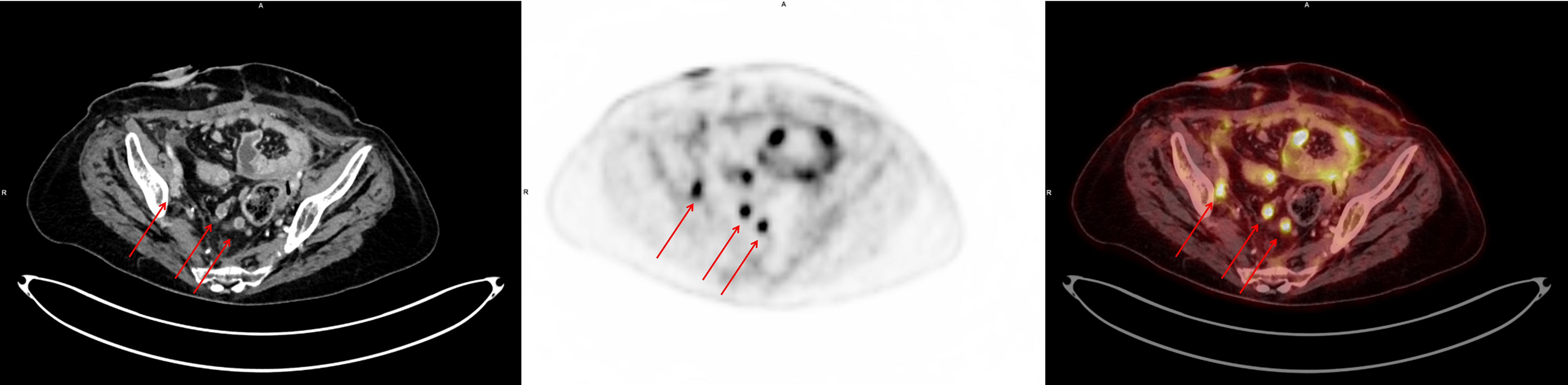
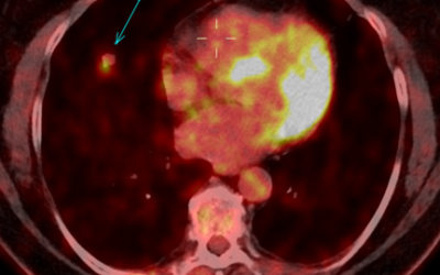
Fig. 1: Exemplary lymph node metastases
Enlarged lymph nodes with high FDG uptake iliac (left arrow, SUVmax 8,2) and pelvic (middle / right arrow, SUVmax 11). Ileostomy in the right lower abdomen.
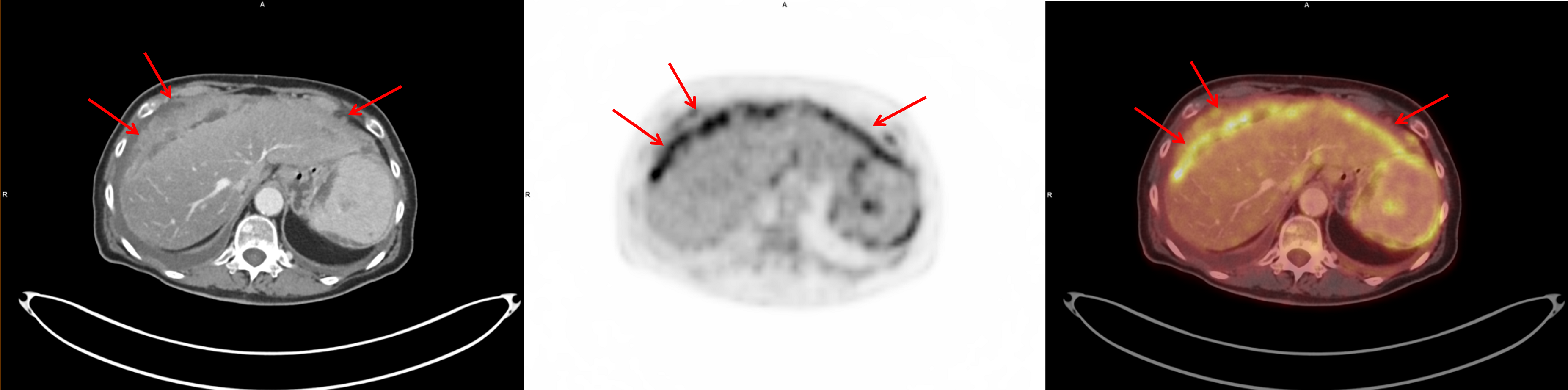
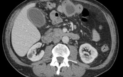
Fig. 2: Peritoneal carcinosis in all four quadrants
Pertioneal tumor mass adjacent to liver and spleen with scalloping and high FDG uptake (SUVmax 9,8). Steatosis hepatis.
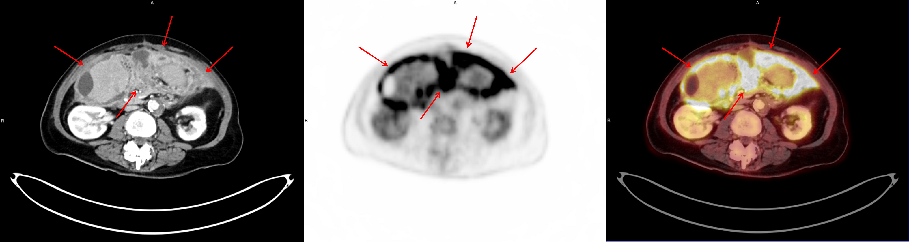
Fig. 3: Peritoneal carcinosis in all four quadrants
Mesenteric and pertioneal tumor mass in the lower quadrants of the abdomen with high FDG uptake (SUVmax 15). Atherosclerosis.
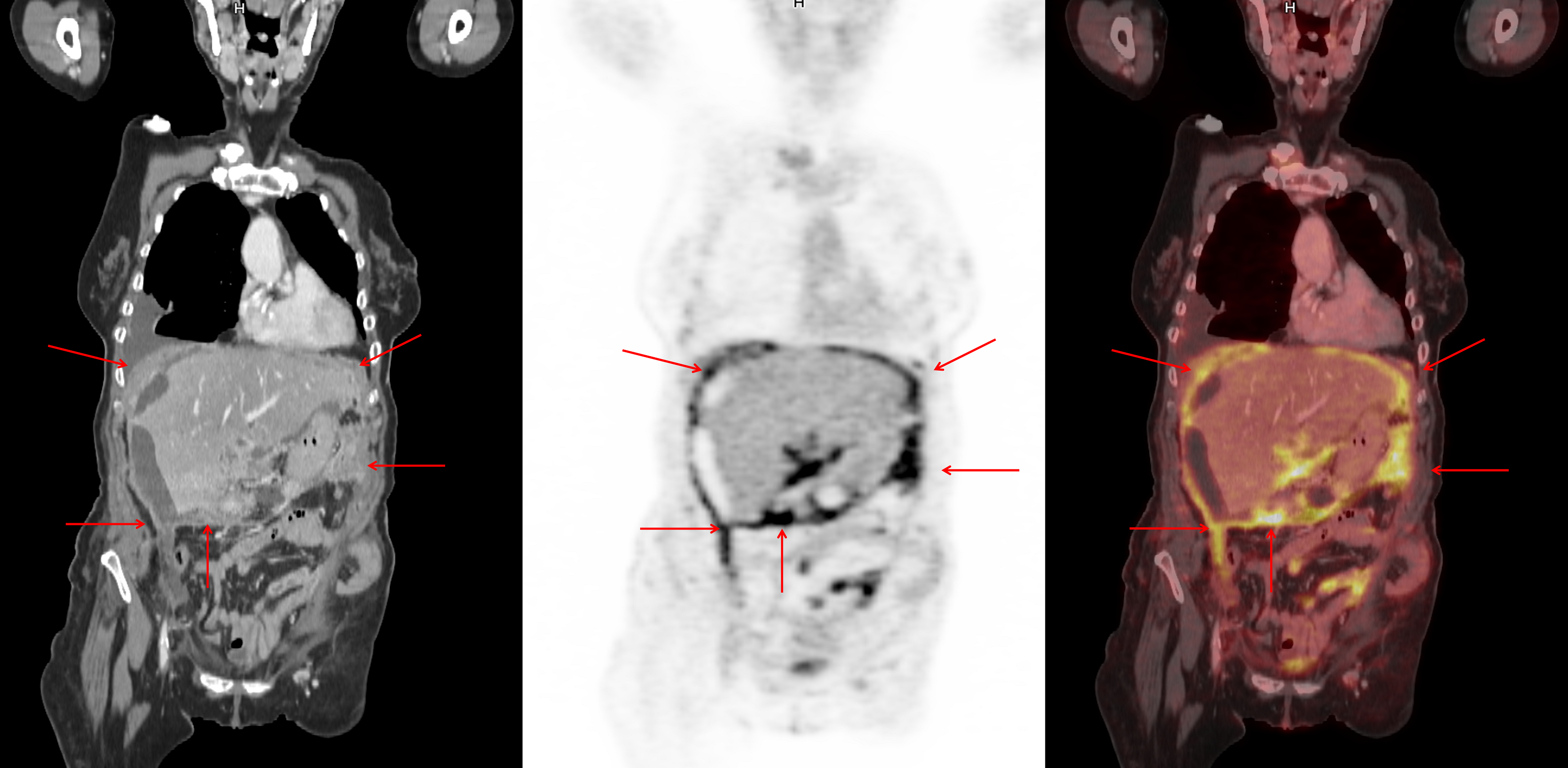
Fig. 4: Peritoneal carcinosis in all four quadrants
Peritoneal tumor mass in all four quadrants of the abdomen with high FDG uptake (SUVmax 15). Right-sided leural effusion.
Description:
Female patient with papillary serous ovarian cancer (PSOC) (pT3c pN1 G3, FIGO IIIC) s/p tumor resection, hysterectomy with double adnexectomy, peritoneal resection, lymph node dissection (pelvic, paraaortocaval, proctosigmoidectomy).
Recent PET/CT scan was performed during chemotherapy (PLD), revealing therapy progress with hematogenous dissemination (pulmonary, hepatic), peritoneal carcinosis in all four quadrants and lymphatic dissemination (cardiophrenic, mediastinal, pelvic, iliac, inguinal), respectively.
This Case was kindly provided by:
Dr. Stella Winter
University Hospitals Munich – Campus Grosshadern
Department of Radiology
Marchioninistrasse 15
81377 Munich
Germany
Contact
More cases:
Case No. 22
Primary intestinal diffuse large B-cell lymphoma (DLBCL)
Case No. 21
Primary carcinoid tumor of the lung
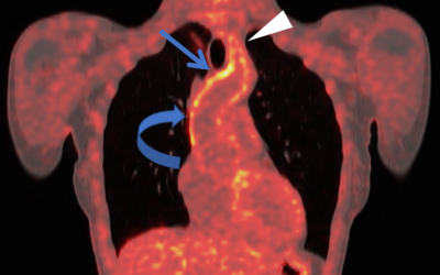
Case No. 20
Takayasu arteritis: 18F-FDG-PET/CT