18F-FDG-PET/CT for oncology:
Detecting secondary cancer
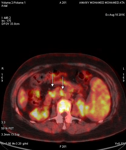
A 51 y/o female s/p lymphadenopathy, splenomegaly and leukocytosis with suspected lymphoma. [18F]FDG-PET/CT was performed in August 2016 for diagnosis and pre-therapy baseline.
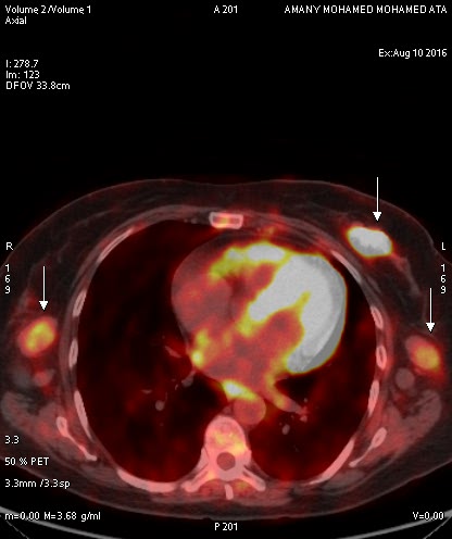
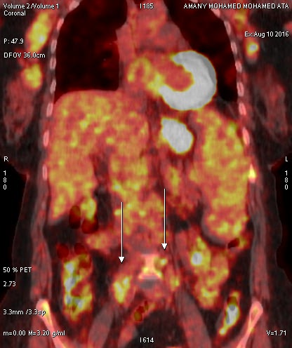
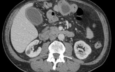
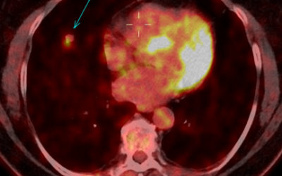
PET/CT study revealed multiple moderately hypermetabolic and enlarged LN’s and multiple splenic and bone marrow infiltrates, highly suspicious of tumour involvement (SUV between 3 and 4). Our study revealed also a glucose avid left breast lesion (involving UIQ) with SUVmax of 15, suspicious of a different pathology – possibly a synchronous 2nd primary breast cancer. (see fig. 1)
Of note, breast involvement in lymphoma patients has been reported in literature. Double Primary Cancers are increasingly seen in clinical routine – thanks to advances in medical imaging, and functional imaging in particular. (see fig. 2)
Subsequent patient management should include image-guided biopsy. Accordingly, a biopsy of the right axillary LN’s and left breast UIQ mass was performed. Histopathology (with IHC) revealed Small B-cell Lymphoma in right axillary LN, while the left breast lesion was an Invasive Ductal Carcinoma (IDC). These findings confirmed our suspicion of Double Primary Cancers.
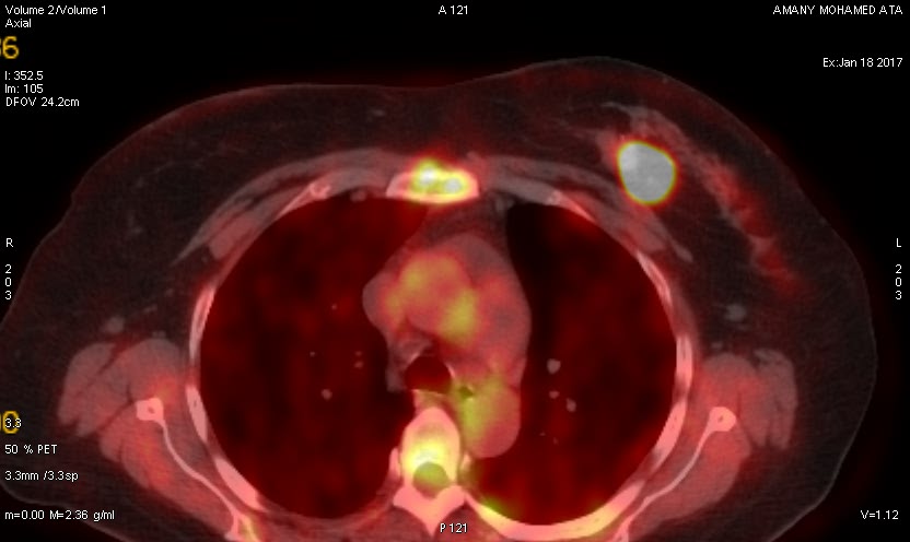
A follow-up [18F] FDG-PET/CT scan was performed after chemotherapy in January 2017, and revealed very favourable therapy response wrt lymphoma with minimal residual nodal disease. In contrast, the left breast cancer (IDC) showed only size reduction with remaining metabolic activity, thus, indicating only partial therapy response.
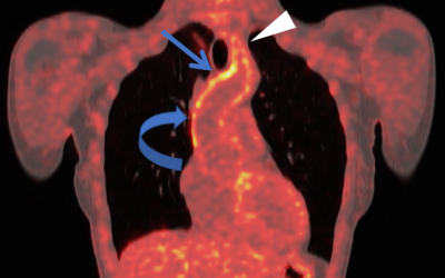
In-conclusion, functional imaging modalities, such as [18F]FDG-PET/CT, are very powerful for the detection, staging, therapy monitoring and follow-up of cancer. Radiologists and nuclear medicine physicians should be able to recognize the spectrum of pathological involvements, common pattern of disease spread, while being asware of alerting observations suggestive of Double Primary Cancers. (see fig. 3)
Breast involvement in lymphoma is a possibility, and, thus, a synchronous 2nd primary breast cancer may be revelealed. In particular, heterogeneous lesion uptake should alert the reader about a secondary tumour, which needs to be confirmed by histopathology.
This Case was kindly provided by:
Prof. Dr. Reda Darweesh
Darweesh Scan Center
39 Sultan Hussein St
Alexandria, Egypt
More Cases:
Case No. 22
Primary intestinal diffuse large B-cell lymphoma (DLBCL)
Case No. 21
Primary carcinoid tumor of the lung
Case No. 20
Takayasu arteritis: 18F-FDG-PET/CT

