[99mTc]-PSMA: xSPECT/CT
Mr WB is a 61 yr old man with a diagnosis of Gleason 9 prostate cancer after a biopsy in May 2017. A subsequent MRI showed a right sided prostate lesion with extracapsular extension, an 8mm left peri-rectal lymph node and right seminal vesical extension. A xSPECT/CT PSMA scan showed abnormal lymph node uptake in paracaval, presacral, left common iliac lymph nodes as well as the right prostate and seminal vesicle.Only in the paracaval group were the size of the nodes increased at 22mm. No bone lesions were identified. His PSA was 20ng/ml at diagnosis. He was subsequently commenced on androgen deprivation therapy and docetaxel in June 2017.
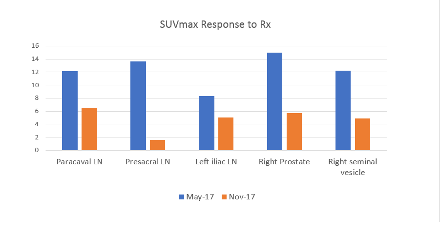
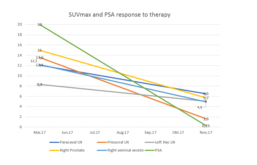
In October 2017, his PSA was 0.25ng/ml. A follow-up PSMA scan was undertaken on 6/11/17 showed reduced uptake and reduced size of all the abnormal lymph nodes (presacral lymph node shown Fig 1), and in the primary prostate lesion (fig 2). Though it is clear that uptake has normalised in the presacral node (Fig 1) there is still persistent uptake in the prostate gland (Fig 2). The latter is thus much better assessed by direct quantification. The measured SUVmax values declined by between 40% and 88% and correlated well with the 99% reduction in PSA (Figure 3). The size of the paracaval lymph nodes declined to 11mm.
This Case was kindly provided by:
 Dr. Iain Duncan
Garran Medical Imaging
2 Garran Place,
Garran, ACT 2605
Australia
garranmedicalimaging.com.au
driainduncan.com.au
Read the story of the GMI here!


