About the centre
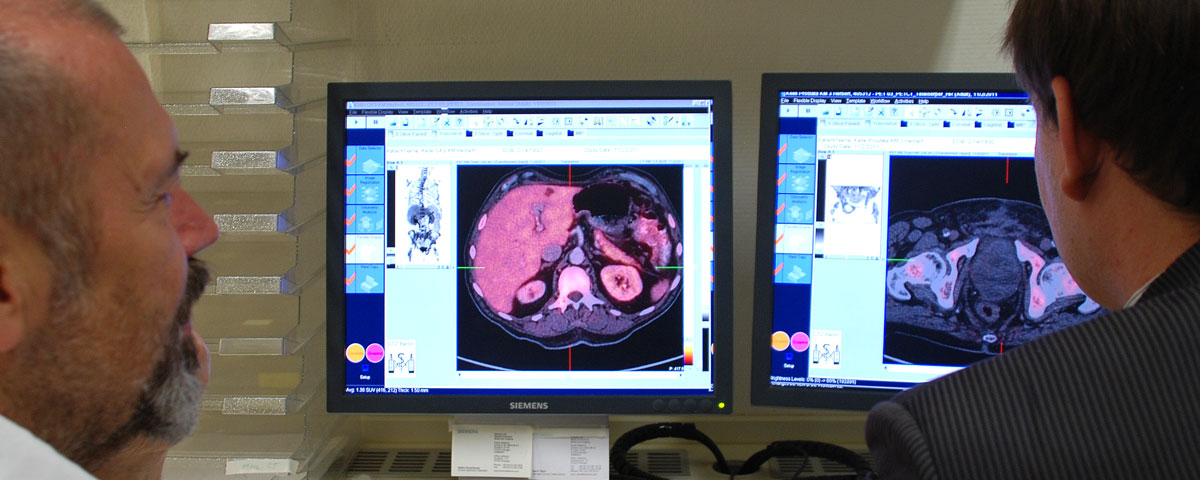
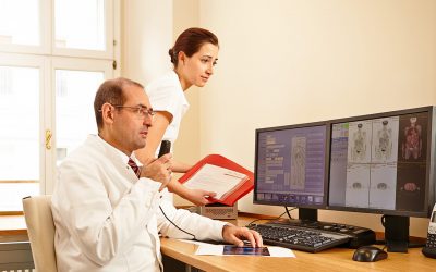
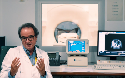
DTZ Berlin has been actively employing PET/CT technology since 2003, which has enabled the centre to offer reliable and precise cancer diagnoses. In 2012, SPECT/CT and MRI (magnetic resonance imaging) were integrated into the hybrid medical imaging program, along with the addition of a modern radiotherapy facility.
Every patient benefits from a highly personalized radiopharmaceutical program using in-house radiochemistry together with a ring accelerator (cyclotron), tested for the highest levels of proven quality.
In 2016, DTZ Berlin added a state-of-the-art PET/MR device to its palette. The program has been designed to unite powerful diagnostic technology with high-performance therapy for comprehensive care tailored to the needs of the individual patient.
Website: www.berlin-dtz.de/en
5 Questions to
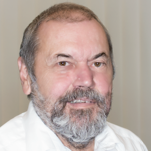
Prof. Dr. Wolfgang Mohnike
 How does the centre work with hybrid imaging?
Our hybrid imaging data flows directly into the process of planning a radiotherapy program, resulting in the best possible treatment. We also work very closely with physicians of various specialties – not only in our own facilities, but also with transferring physicians. Our diagnostics team, comprised of radiologists and nuclear medicine physicians, has collected precise results data using comprehensive state-of-the-art devices, which are used to offer patients more progressive options for continuing therapy.
What is the newest instrument in your program? Why did you choose that one in particular?
Our newest device is a PET/MR system (Biograph mMR). For many different illnesses, the combination of metabolic and anatomical information offers powerful added diagnostic value, which can also play a pivotal role in planning and managing the course of therapy. Moreover, MRI offers a very high soft tissue contrast and a reduction in patient exposure to radiation as compared with CT. This second point is particularly advantageous when it comes to children.
How do the benefits outweigh the costs?
The DTZ has relied on hybrid imaging since 2003 because we are convinced of its diagnostic advantages. We have developed our own in-house certified radiochemistry, which employs its own cyclotron and enables us to cost-effectively create the tracers required for hybrid imaging without sacrificing time or quality. In order to be able to care for our patients in the most flexible way, unlimited by the health politics of the day, we have coordinated specific care provision contracts with several different health insurance companies and participate in numerous studies. Beyond this, we are active in the politics involved in health care in order to promote methods for addressing important issues and the long-term reimbursement of costs for our patients.
How do Radiologists and Nuclear Medicine Physicians collaborate at your medical center?
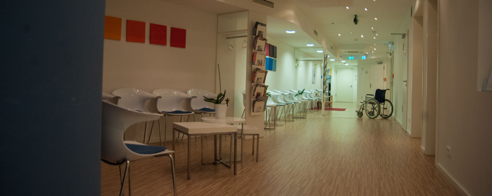
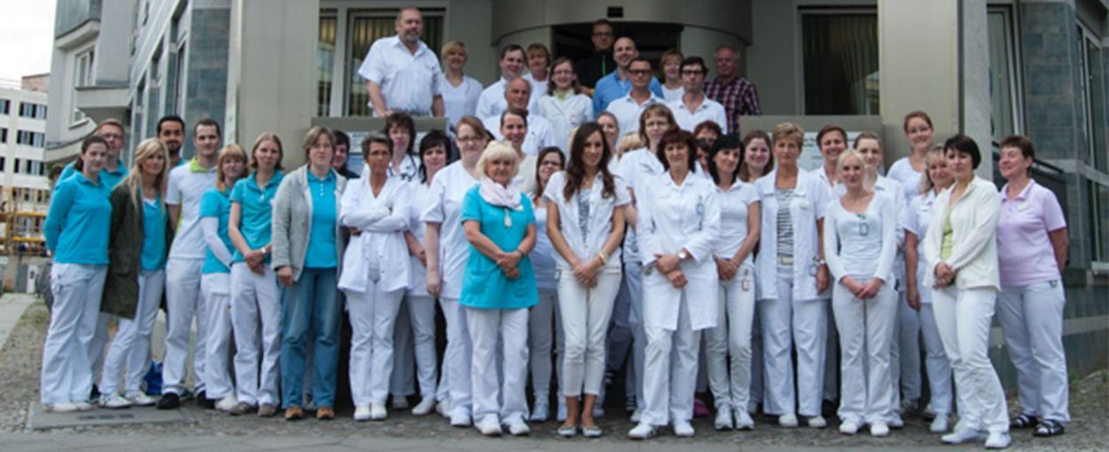
DTZ Berlin has dedicated itself to being an easy-access medical care centre. Our team of radiologists, nuclear medicine physicians and radiologists work together as a single unit under one roof and benefit from each others’ expertise when reviewing patients. This results in the best possible diagnosis and therapy. A communal data pool supports this effective collaboration in that the diagnostic data can be directly applied to therapy planning. Furthermore, many of the physicians possess a dual specialty or are training in a related discipline in order to intensify their knowledge of hybrid imaging.
What will the future hold for hybrid imaging?
In our view, the future of hybrid imaging for nuclear medicine will be determined by the development of new specific tracers under the rubric of “theranostics”, which are suitable for diagnostic and therapeutic application. Highly specific tracers enable not only the confident confirmation or ruling out of illnesses that are difficult to diagnose, (such as F-18-amyloid for Alzheimer’s dementia) or to precisely localize tumours and metastasis that are difficult to detect, but also to treat the corresponding illness (such as Ga-68-PSMA as diagnostic, Lu-177-PSMA as therapeutic tracer for prostate cancer).
Prof. Dr. Wolfgang Mohnike
|
Prof. Dr. Mohnike’sCase to Remeber
|
Would you like to publish your centre on our website and share your story with our community?
Contact: office@eshi-societ.org
More Stories
ESHI Story No. 8
Misr Radiology Center
(Cairo, Egypt)
ESHI Story No.7 (Video)
PETSCAN Vienna
(Vienna, Austria)
ESHI Story No. 6 (Video)
Nuclear Medicine Department / Hospital Sant Pau
Barcelona, Spain
