
About the centre
The Chair of Experimental Molecular Imaging (ExMI) at the Helmholtz-Institute for Biomedical Engineering (HIA) focuses on the development and evaluation of novel imaging methods, contrast agents and theranostics to characterize and treat cancer, cardiovascular and inflammatory disorders. ExMI has a translational scope and many project are located at the interface between preclinical and clinical research. In this context, we often follow a multimodal approach, based on combinations of MRI, CT, ultrasound, optical and photoacoustic imaging, nuclear medicine techniques (in particular PET-MRI) and magnetic particle imaging (MPI). In order to develop image-guided therapies, we strongly interconnect our pathophysiological and pharmacological research with research in device engineering, image reconstruction, and data postprocessing. Basic research on the tissue microenvironment (including the barriers for drug delivery) provides us with new imaging biomarkers and new mechanistic insights that can be used to identify diagnostic biomarkers. Here, imaging features and omics data are considered in concert, evaluated using radiomic and radiogenomic approaches, and used to support mathematical disease models.
As a second main focus area ExMI investigates materials and concepts to improve drug delivery to tumors. This includes the development and evaluation of novel biomaterials, including nanoparticles, polymeric carriers, liposomes, micelles and microbubbles as well as physical and biological treatments of the vasculature and the adjacent tumor stroma in order to improve drug accumulation and tumor penetration.
Currently, the institute consists of 2 departments (Prof. Volkmar Schulz and Prof. Twan Lammers) and 4 research groups working on the biological mechanisms of tumor progression, on novel imaging agents, on medical informatics, on nanomedicines and theranostics, diagnostic and therapeutic ultrasound imaging, and on the development of novel tools for MR-PET hybrid imaging and MPI.
Website: exmi.rwth-aachen.de
5 Questions to
Prof. Dr. Fabian Kiessling
How does the centre work with hybrid imaging?
Our main activities in hybrid imaging include the development of imaging hard- and software as well as in imaging probes for PET-MRI, MPI-MRI, micro-computed tomography – Fluorescence Molecular Tomography (µCT-FMT) and photoaocustic imaging. Furthermore, we apply these technologies in basic research to elucidate biological mechanisms and in translational research to improve patient care.
What is the newest instrument in your program? Why did you choose that one in particular?
The newest devices we acquired were a Philips/Bruker MPI-Scanner and a 7 T MRI (Bruker Biospec) with cryocoils. For the latter, we are currently building a PET-Insert.
How do the benefits outweigh the costs?
We need these devices for our research and assume to translate the money into scientific impact.
How do Radiologists and Nuclear Medicine Physicians collaborate at your medical center?
See description of the institute. In regular meetings between the director of ExMI, the two department heads and the group leaders we discuss and define interesting research topics which we then jointly address. Here we aim for flat hierarchies.
What will the future hold for hybrid imaging?
We see a great future in hybrid imaging as an enabler of precision medicine. In particular, the combination of multiparametric imaging, advanced image processing and radiomics will gain increasing importance in the next years. In this context, we even envision more comprehensive concepts also integrating other diagnostic measures such as omics, clinical chemistry and pathology (see Kiessling 2018 Eur Radiol). In order to implement this idea, the Faculty of Medicine of the RWTH Aachen University has recently founded the Comprehensive Diagnostic Center Aachen, where different medical disciplines, engineers and IT specialists explore innovative diagnostic concepts (for more information visit www.cdca.ukaachen.de).
Kiessling F. The changing face of cancer diagnosis: From computational image analysis to systems biology. Eur Radiol. 2018;28(8):3160-3164.
Prof. Dr. Fabian Kiessling
Since 2008 Prof. Dr. Fabian Kiessling is leading the Institute of Experimental Molecular Imaging at the Helmholtz Institute for Biomedical Engineering at the RWTH-University in Aachen. Fabian Kiessling did his habilitation in experimental radiology in 2006. In 2008 he founded the invivoContrast GmbH together with Matthias Braeutigam.
Fabian Kiessling is author of more than 300 scientific publications and book chapters, edited three books and received many research awards, among those the „Emil Salzer Price for Cancer Research” and the “Richtzenhain Price”.
Fabian Kiessling was in the Editorial board of several scientific journals including Radiology, European Radiology, European Radiology Experimental, and the American Journal of Nuclear Medicine and Molecular Imaging.
He was chairman of the “Molecular Imaging” subcommittee of the European Society for Radiology (ESR), founding member of the European Society for Functional and Molecular Imaging in Radiology (ESMOFIR), is Member at Large in the board of ESHIMT, and treasurer of the European Society for Molecular Imaging (ESMI). Furthermore, he was program chair of the World Molecular Imaging Conference (WMIS) in New York in 2016.
Prof. Dr. Kiessling’s
Case to Remember

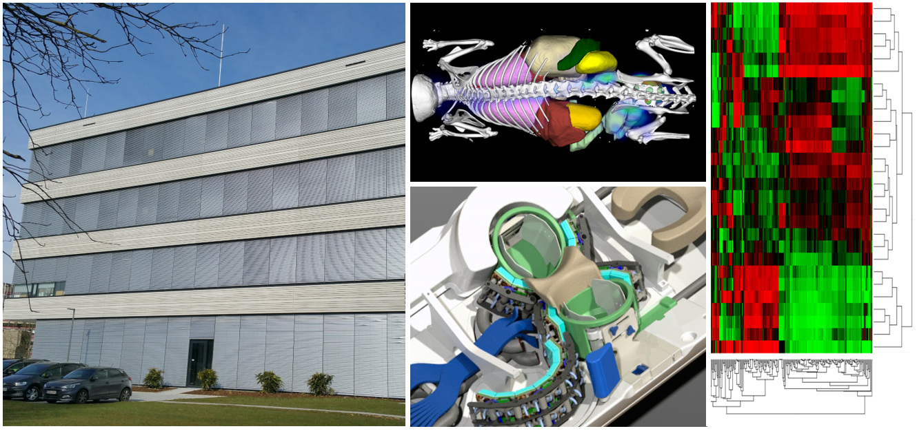
Instrumentation research on simultaneous PET-MRI at ExMI-PMI, headed by Prof. Volkmar Schulz.
Fig. a: PET/MR insert using digital sensor technology for PET;
Fig. b: Derenzo phantom measurements inside and outside MRI showing no distortion of PET and MRI maintaining the submillimeter spatial resolution;
Fig. c: NaF bones scan of a mouse;
Fig. d: simultaneous PET-MRI image of a mouse heart
More Stories
ESHI Story No. 8
Misr Radiology Center
(Cairo, Egypt)
ESHI Story No.7 (Video)
PETSCAN Vienna
(Vienna, Austria)
ESHI Story No. 6 (Video)
Nuclear Medicine Department / Hospital Sant Pau
Barcelona, Spain
Would you like to publish your centre on our website and share your story with our community?
Contact: office@eshi-societ.org