Brain 18F-FDG PET/CT in a child with epilepsy due to Rasmussen encephalopathy

Description:
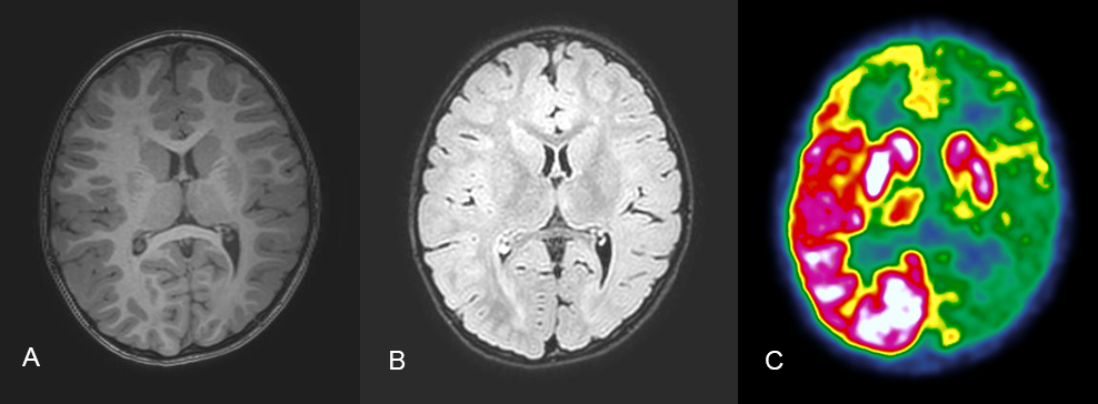
We present a 4 year- old boy with epilepsy since the age of 10 months who had FDG PET/CT scan done in the workup of drug resistant epilepsy. On EEG, a left frontal epileptic activity was identified. MRI showed dysplasia of the left hemisphere of the brain, which was more pronounced in the temporal lobe.
On the PET/CT scan with FDG reduced uptake, almost absent involving extensive areas of the left cortex was found. The differential diagnosis Rasmussen encephalitis (RE), a diagnosis that was biopsy proven.
Discussion:
Rasmussen encephalitis (RE) is a rare, acquired devastating disease that progressively affects one hemisphere. It typically starts in childhood or early adolescence, with a mean age at presentation of 6 years, in a previously healthy child. RE is characterized by brain inflammation with a consequent unilateral brain atrophy. Clinically there are drug-resistant focal or secondarily generalized seizures, worsening unilateral motor deficits, and cognitive decline. The process can be halted only by surgical resection or isolation of the affected hemisphere.
Diagnosis of RE is made based on clinical features and radiological investigations.
This Case was kindly provided by:
Katy Khamis, MD
Department of Nuclear Medicine,
Tel Aviv Sourasky Medical Center,
Tel Aviv, Israel
More Cases
Case No. 22
Primary intestinal diffuse large B-cell lymphoma (DLBCL)
Case No. 21
Primary carcinoid tumor of the lung
Case No. 20
Takayasu arteritis: 18F-FDG-PET/CT